|
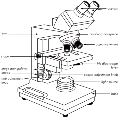
Figure 3.1 Parts of a brightfield microscope. |
|
 |
|
|
|
BIOL 1406
PreLab 3.8
What are the parts of the brightfield microscope?
Cytology is the study of cell structure and function. Because most cells are
very small, they can only be seen when magnified with an instrument such as a
microscope. Microscopes not only provide important information about cell
structure, they also provide clues about how the cell works.
There are 2 basic types of microscopes: the light microscope (creates an image
using a beam of light), and the electron microscope (creates an image using a
beam of electrons.) The most common type of light microscope, and the one you
will use in this lab, is the brightfield microscope. The brightfield microscope
is an example of a compound microscope. That means light from the object you are
viewing passes through two lenses before it reaches your eye.
Microscopes not only magnify the object you are viewing, they also provide
increased resolution. Resolution is the ability to distinguish two points as
separate points. For instance, if two points are very close together, they may
appear to be a single spot. If you increase magnification without increasing
resolution, the single spot will only look like a larger single spot, and will
never resolve into two separate spots. The better the resolution, the sharper
and crisper the image. The resolving power of the naked eye is approximately 0.1
mm, meaning that our eyes can distinguish two points that are 0.1 mm apart. A
light microscope can improve resolution by as much as 1000-fold. In addition,
discernment of cellular detail can be improved with the use of dyes that add
color and contrast to subcellular structures.
|
Magnification of a Compound Microscope |
|
Total magnification = Power of the Ocular lens x Power of the objective lens Example: A typical ocular lens has a magnification of 10x; what is the total magnification if the high power objective (43x) is in position? Total magnification = Ocular (10x) x Objective (43x) = 430x |
In this lab exercise, you will learn about the different parts of the brightfield
microscope, how to care for the microscope, and the correct procedure for
viewing specimens. You will then practice using the microscope to view both a
wet mount and a prepared slide of the organism Euglena.
Study the location and the function of each of the following parts of a
brightfield microscope:
|
Figure 3.1 Parts of a brightfield microscope. |
|
|
|
|
|
|
| YOUR TURN | |
|
The part of the microscope that rests on the table is called the |
|
| A series of lenses that focus light onto the specimen is called the | Check your answer. |
| Which focus adjustment knob should NEVER be used when the high power or oil immersion objectives are in alignment? | Check your answer. |
|
Which part of the microscope regulates the
amount of light passing through the specimen? |
Check your answer. |
| The lenses you look through are called | Check your answer. |
| What is the magnification of the low power objective? | Check your answer. |
|
The surface on which you place your slide is called the? |
Check your answer. |
| The ability to distinguish two points as separate points is called | Check your answer. |
|
Use the drag and drop exercise below to practice identifying the different
parts of the microscope. |
Close this browser window to return to Blackboard and complete the practice quiz and assessment quiz.